 CORE TOPIC:PAGE 3 of 4
CORE TOPIC:PAGE 3 of 4
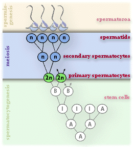
Meiosis
Because prophase of the first meiotic division is prolonged (22 days in human males), primary spermatocytes are commonly seen in sections. At this stage, each chromosome consists of two genetically identical chromatids joined at the centromere. The pairs of homologous chromosomes come into intimate contact with each other and breakages in the DNA strands occur, which result in an exchange of genetic material between the chromosomes. The process is known as crossing over and is an essential feature of meiosis which results in the production of genetically unique gametes.
During metaphase the members of the homologous pair of chromosomes separate, and become attached to a meiotic spindle.
Anaphase follows without delay and the maternal and paternal chromosomes of each pair pass randomly to opposite poles.
Thus at the end of the first telophase, the two spermatocytes contain the haploid number (23) of chromosomes, which is a new combination of maternal and paternal chromosomes.
The second meiotic division follows immediately and no DNA duplication occurs. Because this is a rapid division, secondary spermatocytes are rarely seen in histological sections. The chromosomes of the secondary spermatocytes split at their centromeres in the same way as occurs during mitosis and cell division follows to form spermatids. Spermatids are common in histological sections.
Meiotic division of a primary spermatocyte results in the production of four spermatids which are genetically unique due to the recombination of maternal and paternal chromosomes and the exchange of genetic material at crossing over.
Spermatogenesis
Page 3
<- Page 2|Page 4 ->| Core | Supplementary Material on Spermatogenesis | ||
| Previous Topic Next Topic | Seminiferous tubule (MP) | Spermatogenesis (VHP) | Sertoli cells diagram |
| Sertoli cells (VHP) | Spermatozoa | - | Return to Male Reproductive System Main Index |