 CORE TOPIC:
CORE TOPIC:
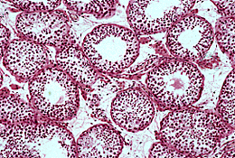
Testis: seminiferous tubules (LP)
Seminiferous tubules
are long, non-branching tubes which open into the accessory duct system through the rete testis. Each seminiferous tubule is 150![]() in diameter and about 80cm long, the total length of seminiferous tubules within each testis is between 300 and 900m. They are very coiled and are seen cut in all planes of section. In a sexually mature male, the tubules have a central lumen surrounded by a complex stratified epithelium and basement membrane. The epithelium consists of two cell types Sertoli cells and spermatogenic cells.
in diameter and about 80cm long, the total length of seminiferous tubules within each testis is between 300 and 900m. They are very coiled and are seen cut in all planes of section. In a sexually mature male, the tubules have a central lumen surrounded by a complex stratified epithelium and basement membrane. The epithelium consists of two cell types Sertoli cells and spermatogenic cells.
Sertoli cells lie between the spermatogenic cells and are important supporting cells. They have several functions including the maintenance of the blood-testis barrier.
Spermatogenic cells undergo extensive proliferatation before undergoing meiotic division. The mitotically active cells lie at the periphery of the tubule, close to the basement membrane. The cells which enter meiosis pass through the epithelium and spermatozoa are eventually shed into the lumen. The spermatozoa are passively transported through the tubules by the secretions of Sertoli cells.
| Core | Supplementary Material on Testis | ||
| Previous Topic Next Topic | Seminiferous tubule (MP) | Interstitial tissue (MP) | Return to Male Reproductive System Main Index |