SUPPLEMENTARY: 2
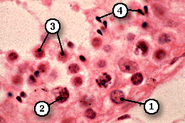
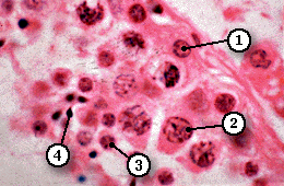
Spermatogenesis (VHP) In these high power pictures the spherical nuclei of spermatogonia (1) can be identified close to the basement membrane. Because prophase of the first division of meiosis is prolonged (22 days in the human male), primary spermatocytes (2) are numerous in any section of seminiferous tublules. They are easy to identify, since they have the largest nuclei with thick, prominent chromosomes.
By contrast, later stages of the first division of meiosis and the secondary spermatocytes of the second division are difficult to identify. Close to the lumen, there are numerous spermatids (3) and the highly condensed, tear-drop shaped nuclei of sperm (4). The cells are closely associatated with Sertoli cells but the arrangement of cell membranes cannot be seen using a light microscope.

