 SUPPLEMENTARY:1
 SUPPLEMENTARY:1
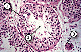
Seminiferous Tubule (MP)
Spermatogonia undergo mitotic proliferation. Most cells enter meiosis but some will remain to form part of a stem cell population which will give rise to further generations of cells.
Primary spermatocytes are common in sections, since prophase of the first division of meiosis is prolonged. They are easy to recognise in histological sections since they have the largest nuclei of any of the germ cell lineage and have obvious, dense chromosomes. Each diploid primary spermatocyte gives rise to two haploid secondary spermatocytes.
Secondary spermatocytes are rarely seen since interphase is very short and secondary spermatocytes pass quickly through the second division of meiosis. Each secondary spermatocyte gives rise to two spermatids.
Spermatids are the most numerous cells of seminiferous epithelium. The cell boundaries are not visible since they lie in deep crypts in the surface of Sertoli cells. They are smaller than spermatocytes and have a high nucleus/cytoplasmic ratio. When spermatids undergo spermiogenesis, they change from a relatively simple, spherical cell to a highly differentiated spermatozoon.
Note that there is no cell division during the extensive reorganisation which takes place as a spermatid forms a spermatozoa. The details of spermiogenesis can be only be appreciated in sections from the electron microscope.
Mature spermatozoa are rarely seen in the tubules because of their rapid transfer to the epididymis.
| Core | Supplementary Material on Spermatogenesis | ||
| Return to Core | Seminiferous tubule (MP) | Spermatogenesis (VHP) | Sertoli cells diagram |
| Sertoli cells (VHP) | Spermatozoa | - | Return to Male Reproductive System Main Index |