 SUPPLEMENTARY: 3
 SUPPLEMENTARY: 3
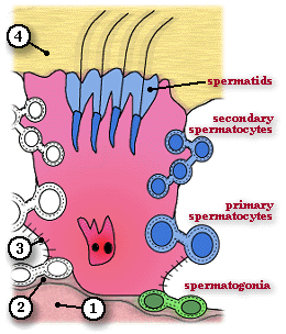
Sertoli Cell Diagram
Since all nutrients must diffuse through the Sertoli cell cytoplasm, these cells control the formation of a micro-environment within the adluminal compartment and the lumen of the seminiferous tubules for meiotic cells and sperm. A particularly important function of the blood-testis barrier is that it prevents the immunological recognition of genetically unique sperm and the possible formation of antisperm antibodies.
The functions of Sertoli cells include:
playing a supporting function by providing nutrients for developing spermatogenic cells
phagocytosing residual cytoplasm during spermiogenesis
producing the testicular fluid which carries sperm through the accessory duct system
producing Androgen Binding Protein which transports testosterone into the duct system and accessory glands (under the influence of FSH)
producting Inhibin (a protein hormone which has a negative feedback effect on FSH release)
producing Mullerian Inhibiting Substance (MIS) during fetal development which inhibits the development of the female duct system
| Core | Supplementary Material on Spermatogenesis | ||
| Return to Core | Seminiferous tubule (MP) | Spermatogenesis (VHP) | Sertoli cells diagram |
| Sertoli cells (VHP) | Spermatozoa | - | Return to Male Reproductive System Main Index |