 SUPPLEMENTARY:
SUPPLEMENTARY:
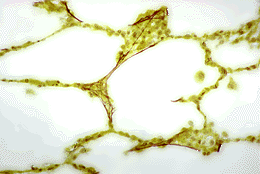
Elastic fibres in the alveolar wall (MP)
This section of lung has been stained with orcein which specifically stains elastic fibres brown.
Elastic fibres contain the protein elastin which has remarkable properties of stretch and recoil. Elastic tissue is abundant throughout the entire respiratory system.
During inspiration, the diaphragm and intercostal muscles contract increasing the volume of the thorax. The resulting decrease in pressure draws air into the lungs and as they inflate, the elastic tissue in the walls stretches to accomodate the inhaled air. Expiration occurs when the intercostal muscles relax, the natural, elastic recoil of the stretched fibres causes the lungs to spring back into shape and causes air to be expelled (i.e. inspiration is an active process whereas expiration is normally passive).
In emphysema, the walls between alveoli are destroyed leading to permanent dilatation of alveolar spaces. The loss of elastic support causes the collapse of bronchioles and leads to obstruction of the airways.