 SUPPLEMENTARY:
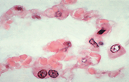
Alveolar wall capilliaries
(VHP)
Examine the walls between the alveoli in this very high power picture of a section of the lung. The extensive network of capillaries in the wall is packed with pink staining
erythrocytes ( (8-10 m in diameter). The capillaries bulge into the alveoli so that a high
proportion of the capillary surface is exposed to the alveolar air.
m in diameter). The capillaries bulge into the alveoli so that a high
proportion of the capillary surface is exposed to the alveolar air.
The wall of the alveoli consists of:
Type I pneumonocytes are squamous epithelial cells which line
97% of the alveolar surface and are in direct contact with the alveolar air.
The cytoplasm of these epithelial cells is so thin that it cannot be seen
with the light microscope. However, the electron microscope demonstrates that the
cytoplasm of these cells forms a complete layer which lines the
alveoli.
Type II pneumonocytes cover the remaining 3% of the alveolar
walls. They have abundant, secretory cytoplasm which causes them to
bulge charactaristically into the alveolar lumen and clearly distinguishes
them from type I cells. Type II cells possess cytoplasmic vesicles
which contain surfactant, a lipid which acts on the the alveolar
epithelium to lower surface tension and prevent the collapse of alveoli
during expiration. There are two type II pneumonocytes at the bottom of this picture.
Elastic fibreselastic fibres provide important support in
the alveolar wall.
Respiratory distress syndrome affects the survival of very
premature babies. They are unable to inflate their lungs due to the great
surface tension between the collapsed walls of the alveoli. They are
treated with artificial surfactant to overcome this problem.
Tutorial Navigator: