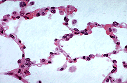
Alveoli are the terminal parts of the bronchial tree and are about 200m in
diameter. Although in this section they appear to be closed sacs, in 3 dimensions they are cup-shaped spaces, arranged like the cells of honeycomb, with an opening on one side which communicates with the alveolar duct. The diagram of the respiratory bronchus illustrates this 3D arrangement which can also be clearly seen in the slide of respiratory bronchioles and alveolar ducts.
The structure of the alveolar wall is specialised to facilitate rapid diffusion of oxygen and carbon dioxide between the blood in the capillary bed and the alveolar air. The alveolar wall between two neighbouring alveoli contains a rich capillary network, sandwiched between squamous epithelial cells supported by a rich matrix of elastic fibres. The detailed structure of the epithelial lining cannot be resolved satisfactorily with the light microscope.
 CORE TOPIC: