 SUPPLEMENTARY:
 SUPPLEMENTARY:
Trachea (MP)
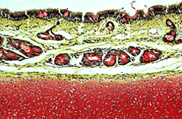
The respiratory epithelium lining the lumen of the trachea, lies at the top of the picture. The nuclei of the epithelial cells are dark staining. Pink stained goblet cells lie between ciliated columnar cells. It is not possible to identify cilia on the luminal surface of the columnar cells at this magnification.
Immediately beneath the epithelium is a yellow stained connective tissue layer, the lamina propria. Although not visible at this magnification, the lamina propria contains a rich capillary bed which helps to warm the air as it passes deeper into the airways. Throughout the entire respiratory system, the lamina propria contains abundant elastic tissue which stretches during inspiration and recoils during expiration. The elastic fibres are not specifically stained in this section.
Pink staining mucous glands lie deep to the lamina propria, in the submucosa. These glands open by ducts on to the surface of the epithelium (there is no duct in this section). The mucus secreted by these glands lies on the surface of the epithelium helping to moisten the air and trapping any particles.
The hyaline cartilage at the bottom of the picture stains pink due to its glycoprotein content. The cartilage rings in the wall of the trachea ensure an open airway at all times.
| Core | Supplementary Material on Trachea | ||
| Return to Core | trachea (MP) | trachea LS (MP ) | tracheal epithelium (HP) | Return to Respiratory System Main Index |