 SUPPLEMENTARY:
  SUPPLEMENTARY:
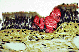
Respiratory epithelium of the trachea (HP)
This is a high power picture of the respiratory epithelium which lines the conducting passages of the respiratory system and the trachea. The epithelium consists of:
Immediately deep to the epithelium is the lamina propria in which there is a rich capillary bed. There are abundant elastic fibres in the lamina propria but they cannot be distinguished by this particular staining method
There is more detailed information about the respiratory epithelium later in this tutorial.
| Core | Supplementary Material on Trachea | ||
| Return to Core | trachea (MP) | trachea LS (MP ) | tracheal epithelium (HP) | Return to Respiratory System Main Index |