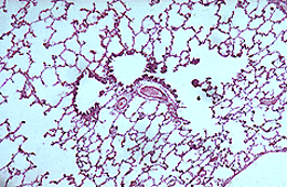
Examine the accompanying photomicrograph of the lung, it consists mainly of sections of alveoli, however, there are two respiratory bronchioles near the centre of the slide. The walls of the bronchioles are interrupted in some places for the openings of alveolar ducts and individual alveoli. In this section, one of the respiratory bronchioles opens into a alveolar ducts. Respiratory bronchioles are lined by a ciliated, cuboidal epithelium. The wall is very thin and consists only of some smooth muscle cells and elastic fibres. An arteriole of the pulmonary artery accompanies each bronchiole.
The alveolar ducts are the last divisions of the respiratory tract to contain any smooth muscle.
 SUPPLEMENTARY: