 SUPPLEMENTARY:
 SUPPLEMENTARY:
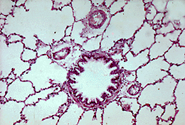
Bronchiole (MP)
This is a section of lung illustrating alveoli and a transverse section through a bronchiole and the accompanying branches of the pulmonary artery. The wall of the bronchiole consists of an epithelial lining surrounded by bundles of smooth muscle fibres and a fibrous coat rich in elastic tissue.
The largest bronchioles are lined by a respiratory epithelium with goblet cells and cilia, but as the bronchioles branch and decrease in diameter, the epithelial cells flatten, goblet cell numbers fall and eventually disappear from terminal bronchioles.
The lamina propria largely consists of smooth muscle cells which contract in response to parasympathetic (vagal) stimulation and relax in response to sympathetic stimulation.
Asthma is caused partly by contraction of bronchiolar smooth muscle which causes bronchoconstriction. Asthma attacks are treated with drugs which mimic the action of sympathetic nervous stimulation. Find out how these drugs are administered and why they are delivered in this way.
| Core | Supplementary Material on Bronchioles | |
| Return to Core | respiratory bronchiole (MP) | respiratory bronchiole | Return to Respiratory System Main Index |