SUPPLEMENTARY: 1
|


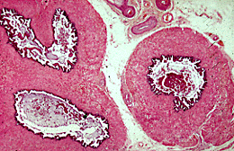
The walls of the seminal vesicles consist of inner and outer layers of smooth muscle and an external layer of connective tissue containing many elastic fibres. The lining epithelium consists of tall columnar cells. The height of the epithelium and its secretory activity are dependant on testosterone secretion.
The epithelium is thrown up into a series of thin folds which increase the surface area of the lining epithelium and allow the organ to distend with secretion. Irregular purple and pink masses of secretion can be seen in the lumen.
The fluid secreted by the seminal vesicles contains abundant fructose, prostaglandins, amino acids, citric acid and ascorbic acid. Fructose is the major metabolic substrate of sperm. At orgasm, in response to sympathetic stimulation, the muscular wall of the seminal vesicles contract to propel the fluid from the lumen of the vesicles into the ejaculatory ducts which are the terminal portions of the vasa deferentia. The right and left ejaculatory ducts pass through the substance of the prostate to join the prostatic urethra at the prostatic utricle.
| Core | Supplementary Material on Accessory Glands | ||
| Return to Core | Seminal vesicle (LP) | Prostatic urethra (LP) | Prostate gland (LP) | Return to Male Reproductive System Main Index |