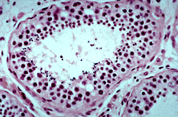
The stratified epithelium which lines the seminferous tubules consists of cells of different sizes. The spermatogenic cells undergo mitotic proliferation followed by meiosis, which results in the production of haploid sperm by a series of events known as spermatogenesis. The diploid cells which give rise to the meiotic cells lie on the basement membrane and as they undergo spermatogenesis, they become intimately associated with Sertoli cell membranes before reaching the lumen where they are released as sperm.
The kinetics of spermatogenesis along the length of a tubule is complex and cells in different parts are at different stages. Examine the epithelium in this picture, in the lower half of the tubule the nuclei of the luminal cells are extremely small and darkly staining, these are the nuclei of sperm. In the upper half of the tubule, the nuclei of the luminal cells are larger and spherical; these are spermatids at an earlier stage of development. The two parts of the tubule are at different phases of spermatogenesis. This staggering of developmental stages ensures that there is a continuous supply of mature sperm.
 SUPPLEMENTARY: 1