 CORE TOPIC:
CORE TOPIC:
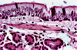
Respiratory Epithelium (HP)
The pseudo-stratified columnar epithelium which lines the conducting portion of the respiratory system consists of a single layer of cells. Some are columnar cells, tall enough to stretch from the basement membrane to the free surface of the epithelium, others are shorter and fail to reach the surface. The shorter cells are basal cells, they are important in the regeneration of the epithelium.
These cell types can be distinguished by their nuclei. Look at the nuclei in the thickness of the epithelium at the top of the picture and notice that:
The respiratory epithelium is modified in two ways to confer very important protective properties on the lining.
Cilia can be seen as a darker band on the surface of the columnar cells in this section. It is not possible to see individual cilia at this magnification (see the electron micrographs).
Goblet cells which produce mucus, lie between the columnar cells and stain darkly purple in this section.
The epithelium is associated with a basement membrane. It forms a boundary between the epithleial cells and the laminia propria but is not specifically stained in this section. The lamina propria is thin and contains numerous longitudinally arranged elastic fibres which have a pink stippled appearance in transverse section. These fibres become stretched in inspiration and recoil during expiration.
Irregular bundles of smooth muscle lie at the boundary of the lamina propria and the submucosa. A group of smooth muscle fibres can be identified in the centre of this picture as a group of pink staining cells with longitudinally arranged nuclei.
At the bottom of this field OUTSIDE the smooth muscle layer, identify serous bronchial glands. Their ducts pierce the smooth muscle and lamina propria to pour secretion on to the surface of the epithelium.
| Core | Supplementary Material on Respiratory Epithelium | ||
| Previous Topic Next Topic | specialisations of respiratory epithelium | goblet cells & cilia | cilia |
| EM of cilia (TS) | pathological increase in goblet cells | metaplasia of the respiratory epithelium | Return to Respiratory System Main Index |